Lesson 1 of 3 in this stage
Evaluating pigmentation: did the colour actually fade?
Separate pigment change from lighting and redness, and evaluate outcomes fairly.
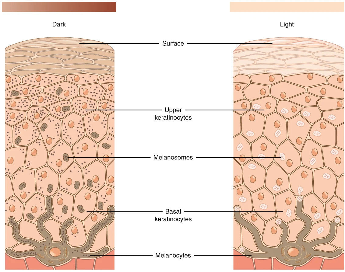
Melanocytes produce pigment and transfer it to surrounding cells. The diagram explains a mechanism; colour alone does not diagnose a dark spot.
Open full-size image ↗Define the feature being followed
Melanin is a natural pigment produced in specialized cells and transferred to other skin cells. Changes in production or distribution can make an area darker than its surroundings. A brown post-spot mark differs from flat redness, and both differ from a depressed scar. Colour measurement alone cannot describe a surface depression. Melasma involves multiple influences and can require a longer, professionally guided plan.
The melanocyte produces melanin and transfers pigment to neighbouring skin cells. Production and distribution both contribute to visible colour.
OpenStax College · CC BY 3.0 · Licence · Open the image to enlargeA photograph can change while the mark does not
Brighter lighting, altered exposure, a filter or moisturizer reflection can make a face appear lighter. Photograph the same area with similar lighting, distance, angle and conditions, without filters. Compare the mark with nearby skin too: a brighter whole image does not prove selective fading. Home photographs are approximate tracking tools, not diagnosis or calibrated measurements.

Find the stratum corneum at the top and cell renewal below. This is thick skin, such as a palm; its clear lucidum layer is absent from facial skin.
Responder counts matter alongside an average
Teaching example: a 20% average improvement could mean similar changes for everyone or large changes in some people and none in others. The average alone cannot distinguish these patterns. Ask about sample size, withdrawals, reasons for leaving and how improvement was calculated. Showing only completers after irritated participants withdraw can make the result look better than the full experience.
Managing the trigger belongs in the outcome plan
If new acne continues, older marks may fade while new marks appear. Track these separately instead of declaring every pigment-focused step a failure. Sun protection and reduced irritation help limit repeated triggers. A new unusual or changing lesion needs medical assessment rather than an unplanned lightening experiment.
Measuring pigmentation: lighting and selection bias
Phone photos vary with illumination and white balance. Colour instruments can quantify defined properties under controlled conditions, but they still require consistent anatomical sites, setup and repeated measurements. Another major bias is selecting the most flattering before-and-after image rather than reporting all pre-specified outcomes.
Interpret a brightening trial
- Determine which pigmentation condition was studied and whether participants share the same underlying cause.
- Check whether sun protection and simultaneous acne treatment could influence results.
- Examine average change, variation between people and follow-up time, not one dramatic photograph.
A lower colour index can be relevant evidence, but it does not by itself establish mechanism or suitability for every kind of dark mark.
Your turn
Use what you learned
In two photographs, the whole face is brighter in the second under stronger light. What is missing for judging a mark?
See the answer and why
Matched photographic conditions, comparison with surrounding skin and tracking the same area. Different lighting prevents isolating the mark's change.
Check your understanding
Answer four questions, then review the reason for each answer. You can retry.